Take Free TSBDE Anesthesia Jurisprudence Practice Exam Now!
In the world of modern dentistry, precision and detailed understanding of a patient’s oral and maxillofacial structures are key to delivering effective treatment plans.
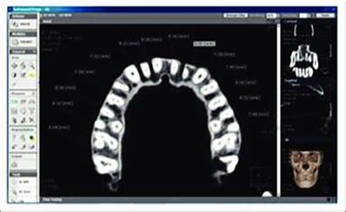
As technology continues to evolve, the use of sophisticated tools like Cone Beam Computed Tomography (CBCT) has become a standard in dental practices worldwide. CBCT, originally developed for use in dentistry in the mid-1990s, allows for accurate three-dimensional scanning of osseous or bony structures within the oral and maxillofacial region.
These scans generate various file formats that carry a wealth of information, integral to dental diagnosis and treatment planning. In scenarios such as implant planning, endodontics, maxillofacial surgery, and orthodontics, traditional 2D images often fall short of providing the necessary depth of information. That’s where CBCT comes in, filling the gap and enhancing diagnostic accuracy and precision.
In this article, we take a closer look at how CBCT files contribute to dental diagnosis and treatment planning and shed light on the different file formats produced by a CBCT scan, underscoring their role in revolutionizing dental care.
CBCT files facilitate surgical planning for procedures such as wisdom tooth extractions, bone grafting, orthognathic surgery, and temporomandibular joint disorder treatment.
Implant Dentistry
CBCT scans enable precise implant planning by assessing bone quality and quantity, identifying vital anatomical structures (such as nerves and blood vessels), and determining the optimal implant size, angulation, and position.
Endodontics
CBCT files help in the management of complex endodontic cases, such as identifying additional canals, detecting root fractures, and assessing the root canal anatomy.
CBCT File Formats in Dentistry
Like 3D printing and CAD/CAM dentistry, CBCT scans can produce various file formats:
- Digital Imaging and Communications In Medicine (DICOM)
- Tagged Image File Format (TIFF)
- Bitmap Image Files (BMP)
- Joint Photographic Experts Group (JPEG)
DICOM (Digital Imaging and Communications in Medicine)
DICOM or DCM, Digital Imaging and Communications In Medicine file, is known to be the universal file format for 3D CBCT images, both in medical imaging and in dental CBCT. Not only does this format store image data but also relevant patient information and scan parameters for accurate diagnosis and treatment planning.